
Sponsored by Beekley Medical
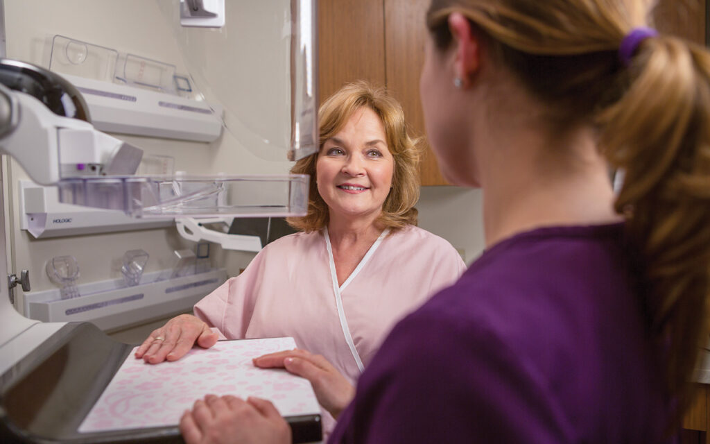
 Women’s imaging is one of the most dynamic and evolving areas in healthcare, with advances in technology, patient-centered care, and population health initiatives shaping its future. Radiology and imaging directors are at the forefront of these changes, working to expand access, improve outcomes, and enhance the patient experience.
Imaging Community Exchange (ICE) Magazine invited leaders in the field to share their perspectives on the opportunities, challenges, and trends that are shaping women’s imaging today.
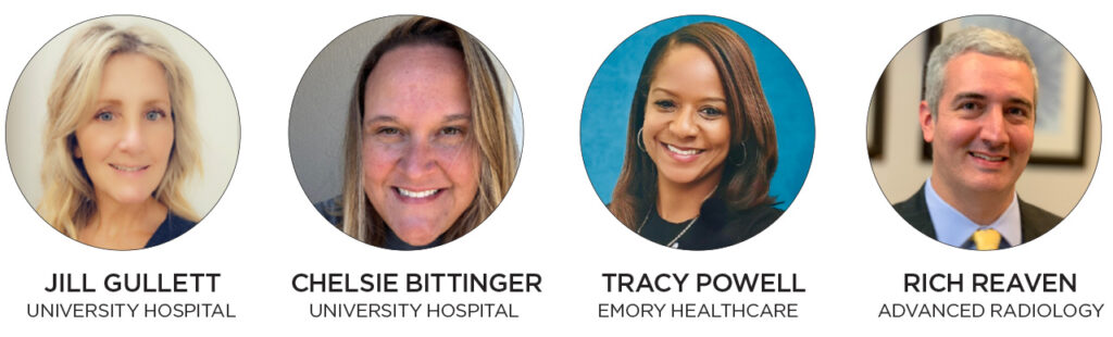
Participants in this Director’s Circle article on women’s imaging are:
- University Hospitals Breast Center Assistant Nurse Manager-Radiology Department Chelsie Bittinger, RN, BSN, CBCN;
- University Hospitals Breast Centers Mammography Supervisor-Radiology Department Jill M. Gullett, RT(R)(M); and
- Emory Healthcare Director of Patient Care Services-Radiology Tracy E. Powell MSN, ANP-BC;
- Advanced Radiology’s Richard Reaven, MD.
Bittinger and Gullett combined their answers. Their answers are labeled as B&G.
Q: Women’s imaging continues to evolve with advances in technology and screening guidelines. From your perspective, what is the biggest opportunity right now to improve outcomes for patients?
B&G: The biggest opportunity to improve outcomes in women’s imaging can be by expanding access to care, providing faster results and clearer communication, using advances such as abbreviated MRI, contrast-enhanced mammography and applying AI tools to ensure consistent quality promotes better outcomes. By reducing anxiety through timely access, quick results, and shared decision-making, we can help patients stay on track with care and improved outcomes. With innovative approaches like abbreviated MRI, contrast-enhanced mammography, and AI tools, we can expand access to high-quality imaging and increase adherence to screening guidelines.
POWELL: The biggest opportunity for women’s imaging is increased education around the importance of early screening and detection. Daily, radiologists play a significant role in identifying abnormalities, especially with mammography. We now utilize digital breast tomosynthesis or 3D mammography which improves cancer detection. Also, interventional radiologist have various treatment options for pelvic health such as treatment for pelvic venous congestion and uterine fibroids.
REAVEN: I think the biggest opportunity to improve outcomes will be to address the current struggles getting women to come in for the annual breast imaging. Right now, only about half of the women over age 40 currently get an annual breast imaging examination. The other half are still experiencing breast cancer, we just aren’t able to find it if we can’t see it. Our job as breast imaging radiologists is to find and diagnose breast cancer at its earliest and most treatable stage. One of the ways that will be important moving forward will be compression-free options that are just as accurate or even more accurate than a mammogram, including dedicated low-dose breast CT device.
Q: How is your department addressing disparities in access to women’s imaging services, particularly for underserved or rural populations?
B&G: Imaging departments can reduce disparities and improve access to women’s imaging services by combining technology, community engagement, and practical support. Strategies include scheduling teams offering incentives to rural patient populations, deploying mobile mammography units to bring care directly to underserved areas, and partnering with local organizations to provide outreach and education on breast health and screening guidelines. These efforts make imaging more convenient, raise awareness, and ensure essential services reach all populations.
POWELL: The disparities in women’s health remain a huge issue when so many women are underinsured. This particularly affects women of color and those living in rural areas. The only way to close the gap is through private and federal funding of programs focused on preventative care and early detection. We must also address the shortage of physicians in rural areas. Care can be expanded with increased utilization of advanced practice providers such as NPs and PAs.
REAVEN: Mobile imaging units can play a significant role in rural populations, and hopefully local county and state specific health outreach programs can help the underserved populations. We obviously need more of both of these in order to make a significant impact in underserved and rural women’s health.
Q: Artificial intelligence and advanced imaging technologies are increasingly integrated into breast and pelvic imaging. How are you balancing innovation with accuracy, patient safety, and workflow efficiency?
B&G: To effectively leverage AI and advanced imaging technologies, departments must thoroughly validate tools for accuracy, maintain ongoing quality control, and integrate them seamlessly into existing workflows. Continuous staff training helps radiologists, technologists, and support teams understand both the capabilities and limitations of these innovations, reducing errors and enhancing proper use. Prioritizing patient safety through system safeguards and protocols, along with streamlined workflows to prevent bottlenecks, ensures reliable and efficient care. Ultimately, careful evaluation of these innovations, well trained staff, prioritizing patient safety measures, and adherence to ethical standards are essential for maximizing the benefits of these technologies.
POWELL: There must be checks and balances when using modern technology to ensure accuracy and patient safety. One way would be to use AI as a second reader tool and not a replacement for human beings. Also, regular audits would be essential to track discrepancies between AI and radiologists.
REAVEN: I think we need to carefully weigh the pros and cons of artificial intelligence before we go straight into full deployment across the board. There are a lot of questions that remain to be answered, including: Who will pay for these technologies? What are the medicolegal implications of using AI in the radiology space if there is an explicit plan to replace the human radiologist?
Q: What strategies have been most effective in improving patient experience and reducing anxiety during mammography, ultrasound, or MRI for women’s health?
B&G: Creating a positive patient experience in women’s imaging requires efficient scheduling, clear communication, supportive staff, and a focus on privacy and dignity. Reducing wait times through streamlined workflows helps minimize anxiety, while offering calming options such as music or visuals can further ease stress. Encouraging questions and involving patients in care decisions enhances their sense of control and comfort. Together, these strategies build trust, reduce anxiety, and create a more supportive environment throughout the imaging process.
POWELL: The best strategy for any examination is the human connection with empathy and communication. Using technology such as videos to view prior to testing along with instructions. For mammography especially, taking time to explain the imaging test prior and being honest that the test is not comfortable but using reassurance will help alleviate anxiety.
REAVEN: Education about what to expect is the most important strategy in reducing anxiety and discomfort. But again, using a compression-free imaging modality can be extremely helpful in reducing barriers to entry.
Q: Looking ahead, what do you believe will be the most impactful trend or development in women’s imaging over the next five years?
B&G: Over the next five years, women’s imaging will be reshaped by artificial intelligence (AI), machine learning, and modern technologies like contrast-enhanced mammography (CEM). AI will improve early detection by spotting subtle changes on mammograms, ultrasounds, and MRIs that can lead to faster and more accurate diagnoses. Risk-based screening tools will allow more personalized schedules, while AI-powered education and communication can reduce anxiety and keep patients more engaged. Automation of routine tasks – like triage, lesion characterization, and reporting – will ease radiologists’ workload and speed up results. CEM, which uses a contrast agent to highlight abnormal areas, provides a cost-effective alternative to breast MRI – especially for women with dense breasts or higher cancer risk. Together, these advances will make women’s imaging more precise, efficient, and patient-centered.
POWELL: The advancements in using pelvic MR for quicker diagnosis of chronic pelvic pain. Also, mammography advances such as contrast enhanced mammography or whole breast ultrasound for dense tissue will be impactful. Finally, robust education on preventative healthcare and early detection. This is a moot point if healthcare is not affordable or accessible. Reducing the number of uninsured Americans is essential.
REAVEN: I think that finding the answers to the questions above will be most impactful in what transpires over the next few years. How we decide what role AI plays in the radiology workflow will be essential. Also, finding a way to bring more women into an annual breast imaging examination will be essential.
For more information about Bleekley Medical call 1-800-233-5539, visit our website at beekley.com, or email info@beekley.com
