
 By Matt Skoufalos
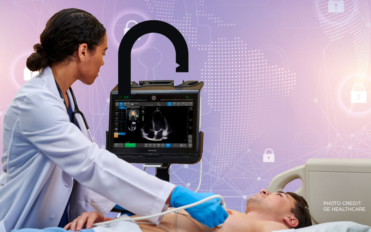
With study volumes continuing to climb – along with wait times in patient scheduling and staffing shortfalls that don’t show signs of self-correction – health systems are seeking more and different ways to increase access to medical imaging services without dramatically elevating costs. In the past decade-plus, diagnostic ultrasound has emerged as a fairly ubiquitous, low-barrier option with an increasing number of use cases. As the medical imaging industry works to address its capacity issues, practitioners are finding that ultrasound represents a versatile first-read option for more and different care environments.

Kassa Darge, radiologist-in-chief and chair at Children’s Hospital of Philadelphia (CHOP) and a professor of radiology and surgery at the Perelman School of Medicine at the University of Pennsylvania, said ultrasound technology is particularly suited to pediatric patients in ways that other modalities may not be.
For a start, it helps parents and practitioners avoid exposing their children to the ionizing radiation of computed tomography (CT) scanners, the requirement to hold a specific position that magnetic resonance imaging (MRI) demands, or exposure to sedation or anesthesia as a condition of such studies. Unlike either of those modalities, parents can sit with their children for an ultrasound, and even help to position them under the guidance of a sonographer. Pediatric institutions like CHOP even warm the transducer gel before applying it to the patient for an added level of comfort, Darge said — a step that’s not typically taken in caring for adult patients.
When a study does call for the use of contrast agents, recent implementation of contrast-enhanced ultrasound has mitigated the potential of contrast adverse events in children, and is even safe to be used in patients with reduced kidney function.
“It has opened new doors for ultrasound that make it possible to diagnose many issues without MR and CT, sedation, radiation and less safer contrast agents,” Darge said. “Additional advanced ultrasound modalities enhance our possibilities, including being able to measure organ stiffness or fat content; things that were done with MRI before.”
In addition to those applications, computing processes powered by artificial intelligence (AI) support automated organ measurement and volumetric calculations, as well as workflow enhancements that improve repetitive processes and make them more user-friendly, Darge said. These technologies deliver a competitive edge for ultrasound in terms of diagnostic capability, cost and accessibility, and can help maximize its potential in a variety of use cases.
“For pediatrics, this enhancement is not just measuring an organ, but making the study faster – which, in the case of a restless child, is really a positive,” he said. “Some manufacturers have added pathology-recognizing AI applications; having such an application for a child that’s crying and moving is a huge additional advantage.”
Tele-ultrasound can offer new options for practices that are expanding. CHOP’s pediatric radiology services are available at 12 sites in New Jersey and Pennsylvania, with future expansions planned into Delaware. Additional ultrasound studies from those remote sites hit the workflow queues at CHOP, and there’s less oversight over how they’re conducted from the centralized radiology department.
The promise of tele-ultrasound would allow radiologists to see the images in real-time during the scan, and even speak directly with the sonographer – advantages that can be consolidated further with the addition of a robotic scanner arm that can be directed remotely by an offsite practitioner. These functions could support multisite radiology operations in a way that that will reduce the impact of the skilled labor shortage in the imaging field at present.
“It’s very hard to put a pediatric radiologist in every satellite site where there are pediatric patients,” Darge said, “but you do need experienced personnel; the machine will not do it all alone.”
Point-of-care ultrasound (POCUS) remains a growth area throughout the field, including at CHOP, allowing for bedside imaging without the need for a large, wired, scanning device. Its continued advancement, which is being driven at the point of clinical education from whitecoat ceremonies to specialized sonography training, allows patients to get their initial ultrasounds “right where they need it,” Darge said, whether for procedural or diagnostic purposes.
This is especially so in a global context, where physicians across the planet have leveraged POCUS to overcome challenges of access, staffing and delivery of results.
“In low-resource countries, the impact of ultrasound is much more significant,” Darge said. “Access to CT and MR is very limited, so radiologists maximize the use of ultrasound. That’s something we should not forget: every advancement of ultrasound that we do here has a global impact.”
 Hannah Simmons is director of product for Inteleos, the non-profit organization that oversees the American Registry for Diagnostic Medical Sonography (ARDMS) the Alliance for Physician Certification & Advancement (APCA), and the Point-of-Care Ultrasound Certification Academy.
Simmons said that her organization sees a lot of global growth for POCUS in less-regulated, less-standardized spaces. The challenge from there remains finding the best ways to integrate this newer tool within established institutional and societal protocols.
“As we have more people using ultrasound, clear competency expectations can help translate training into practice as hospitals adapt workflows to support broader ultrasound use. It’s one thing to incorporate into sonography training programs, but it’s quite another thing to have hospitals shift their workflows to accommodate these skills that practitioners are acquiring,” Simmons said.
“We can provide this up-skilled certificate program, but it’s not as important to practitioners to do this in a formal way until it’s going to be able to translate in their day-to-day practice,” she said.
Adding contrast-enhanced ultrasound to the national sonography curriculum is one example of the industry finding ways to build processes that support existing use cases, Simmons said. Most hospitals can offer contrast-enhanced ultrasound, but break up the responsibilities for administering contrast agents, capturing images and reading studies across multiple professionals’ roles. Advanced credentialing must accompany meaningful changes to adjusted scopes of practice in day-to-day operations to help maximize patient access to such services.
Some of these shortfalls can be overcome by technology, but Simmons pointed out that many of them can be addressed interpersonally, too.
“Hands-on training is easy to come by in hospitals, and that’s what we find a lot of people are doing,” she said. “AI support is still evolving, but technology in general helps to make it so that we don’t need the expert right beside you. You could message your mentor, your colleague, while out in the field; mentor-mentee relationships help to close that diagnostic gap with complicated cases.”
Along with the expanded access to services that POCUS and broader adoption of ultrasound applications provides, healthcare institutions must also help provide the capacity to support action on study findings. These challenges may be more pronounced in rural and distributed areas, where, if a study identifies something that must be resolved, the institution must have the capacity to address it.
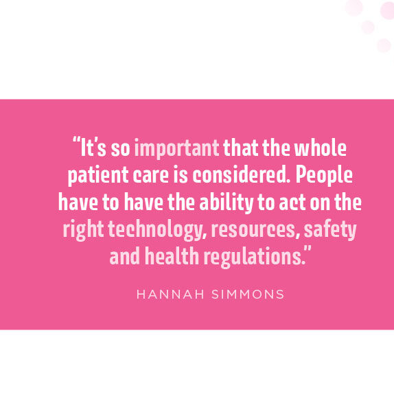
“You need to make sure that you have the capacity, the resources, even the workflow in place to act on that,” Simmons said. “It’s so important that the whole patient care is considered. People have to have the ability to act on the right technology, resources, safety and health regulations.”
Patient access to ultrasound will always be contingent upon practitioners’ access to meaningful, standards-based training, and in the push to ramp up trained sonographers to address increased study volumes, Inteleos is working to deliver supplemental training through virtual reality (VR) and augmented reality (AR) education.
“It sounds counter-intuitive, but if we want to continue to provide good, strong education, and people aren’t seeing certain cases, this is a way to get more people trained in ultrasound,” Simmons said. “We’re not only using AI and new technologies for procedural assistance, but also to make education and training more scalable, so more practitioners or students are able to see different types of things in the field.
 “We all care about patient safety,” she said. “That peer-to-peer mindset gets increasingly more important as we learn things here around the globe. Making sure that our findings are shared globally is so important; connecting people with regional health hubs across the world. The more partners we can have to improve patient care, the better it will be.”
As president and chief commercial officer of ultrasound probe manufacturer Vave Health of San Jose, California, Timothy Kartisek is bullish on the growth potential of POCUS, particularly when paired with AI-powered technologies and in global market settings.
Since its soft launch in the spring of 2025, Vave Health has sought to make inroads at some of the lowest levels of direct patient care with its universal probe. Kartisek described prospective customers in emergency medical services (EMS), sports medicine, rural healthcare, and sub-acute settings, all the way up to medical-surgical areas and frontline medicine in developing markets.
“Our organization was designed to create a device that offers a universal approach to imaging: whole-body ultrasound,” Kartisek said. “We’re using transducers made from PZT crystals, and designing this product to be used in the front lines of healthcare.”
Companies like Vave are looking to compete in the spaces between those filled by larger competitors by building an ultrasound device that doesn’t rely upon wired networks, cart-based systems or physicians’ credentials. It’s designed to be high-quality but low-cost, “scalable without adding a ton of friction to the places it’s being deployed,” Kartisek said.
“We’re excited that more and more people are getting trained on how to use this technology to provide a front-line level of care,” Kartisek said. “We’re creating very specialized workflows dictated to us from the providers. We’re talking to users on the front lines of Africa, Asia, on the battlefield – places that are not typically a customary spot for ultrasound to be done – and layering AI on top of it.”
Kartisek wants Vave to help practitioners to “deliver a first look through ultrasound,” addressing “speed to care” through AI-managed data sets that source study data across health systems to help build workflows that trigger protocols based upon users’ real-time results.
“We’re increasing throughput because we’re able to scale patients, rank the levels of how quickly they need care, and drive down costs,” he said. “We’re really trying to help the health system, help the provider, learn what’s going on with the patient so they can discharge the patient or get them care quicker.”
“The winners in this are not going to be the people with the flashiest demo or the biggest number of sales reps out there,” Kartisek said. “They’re going to be the guys that can take POCUS and make it deployable at scale inside health systems and ministries of health around the world at the lowest cost possible. Our goal is that 10 years from now, we have advanced POCUS so far that if you see a monitor in a room that provides patient care, you see an ultrasound device bolted to the side of it.”
Jocelyne Mekontso, head of diagnostic ultrasound at Siemens Healthineers, said that her company has been most focused on closing gaps in access to diagnostic imaging by automating mundane operational tasks with practical AI. Such technologies can improve conditions for clinicians that frees them up to focus on patients’ needs, whether by reducing operators’ hand movements to cut back on stress-related injuries and minimize repetitive tasks, or by automating workflow to improve the speed and ease of exams.
In addition to serving clinicians, however, diagnostic imaging innovations continue to represent a vital component of improved patient care, Mekontso said. Advanced ultrasound solutions particularly support increased patient access to care not only through diagnostic applications, but also through interventional means, whether guiding biopsies, delivering other therapies or supporting follow-up care.
“I can now see diagnostic treatment in the middle of intervention,” she said. “If we can shorten that cycle for patients, make sure we provide the right care, I think ultrasound will allow us to really be part of the full continuum of care for the patient, depending on the disease state that the patient is being evaluated for.”
Another major policy change supporting expanded use of contrast-enhanced ultrasound – which Mekontso said she’s anticipated for two decades – involves the recent doubling of the CMS reimbursement rate for non-cardiac, contrast-enhanced ultrasound.
“This is also where the policymakers are seeing how beneficial ultrasound could be,” she said; “how economically viable for institutions and also the patient.”
Most of all, ultrasound — like any imaging modality — is only as significant as the ways in which it’s deployed, and the skill with which its operator employs it, Mekontso said.
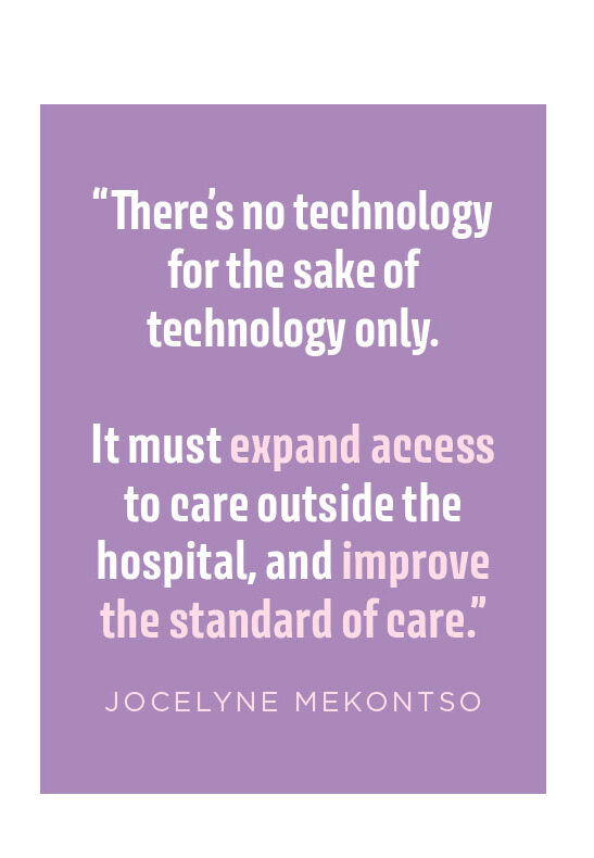
“There’s no technology for the sake of technology only,” she said. “It must expand access to care outside the hospital, and improve the standard of care.”
