
By Matt Skoufalos
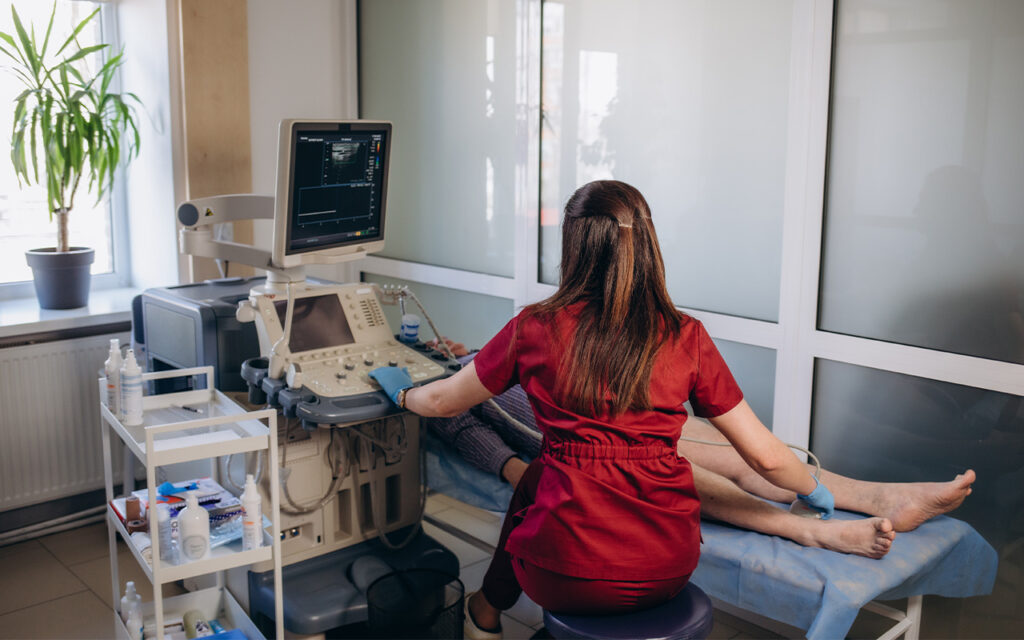
Since its emergence subsequent to the Gulf War of 1990, point-of-care ultrasound (POCUS) has been leveraged in broader and broader use cases in various healthcare settings. Portable ultrasound technology, which sprang from a U.S. Department of Defense (DOD) Defense Advanced Research Projects Agency (DARPA) challenge grant for the development of a diagnostic technology that could reach soldiers in the battlefield during combat, led to the creation of the first widely produced portable ultrasound device, the Sonosite 180.
Coupled with the emergence of the FAST exam (Focused Assessment with Sonography for Trauma), which was developed by trauma surgeon Gail Rozycki, the first, cart-based, portable ultrasound systems began to find practical use in civilian healthcare environments.
Rob Ferre, MD, the POCUS program director for Indiana University School of Medicine and the system-wide POCUS director for the Indiana University Health System in Indianapolis, Indiana, marks that inflection point as the change that began taking diagnostic ultrasound technology from the imaging suite into the emergency department.
“Back then the machines were huge, and they never moved,” Ferre said, “but that new, highly portable form factor really started moving ultrasound into the emergency department.”
In 2001, the American College of Emergency Physicians released its first guidelines for the use of ultrasound imaging in the emergency department. Thereafter, when other specialties from pulmonary critical care to hospital medicine to anesthesia began leveraging the technology, the term POCUS was coined. In 2023, the Accreditation Council for Graduate Medical Education (ACGME) named POCUS a requirement of residency training in family medicine, and now more than half of all medical schools have some approved POCUS curriculum, Ferre said.
“It’s become a bigger deal,” he said.
Ferre traces the ubiquity of POCUS to the development of ever-smaller ultrasound devices, specifically, the Butterfly iQ, which lowered the cost for a handheld portable ultrasound scanner to a few thousand dollars. That was a game-changer for educators.
“Just like when Sonosite 180 came along in the ’90s, Butterfly took the ability to do mass training to the next level,” he said. “Now that POCUS was affordable, how do we develop more experts in the field, and how do we find space in the curriculum for instruction?”
By distributing POCUS systems at white-coat ceremonies, as has become popular to do at medical schools across the country, physicians-in-training now have the opportunity to pick up the technology as they learn anatomy, which offers a practical tie-in for both modes of learning.
“They’re learning to use a modality that’s in clinical practice and for physical exams,” Ferre said. “Now, because we are in a new paradigm where we’re trying to get students to use an imaging modality that’s used in clinical practice, we’re getting them to use this, and we’re making it cheap and available throughout medicine.”
Adding ultrasound technology at the point of education has led to the emergence of a grassroots group of students who are looking to then learn how to incorporate the technology into clinical practice, Ferre said. He believes that the addition of POCUS into such curricula has even become a signifier to medical students that they’re at an institution that embraces innovation.
Beyond that, Ferre spoke about the work that POCUS manufacturers have done to “remove the proximity to education” for training new users of the technology. By adding in access to diagnostic assistance tools and artificial intelligence (AI)-powered image recognition algorithms, device-makers are “helping the new user, and giving the user who feels uncomfortable an added boost” to embracing POCUS.
“I call it ‘just-in-time learning,’” Ferre said.
“As I’m using the device, can I quickly refer to some educational materials? If I want to refresh my memory, how can I do it?” he added. “It helps overcome some of the reluctance to adopt the technology.”
Those same utilities also make POCUS a technology ideally suited for use by “downstream healthcare workers” who might not have the same levels of medical training as a physician or modality specialist, but who can quickly pick up specific use cases for which POCUS is particularly suited, Ferre said.
“We know that vascular access is difficult,” he said. “If I can give a nurse or phlebotomist a technology that can help them perform the procedure more easily, then why shouldn’t I? Lung ultrasound is another one of those very good, easy-to-learn applications that has a good use case. It’s just pattern recognition, and you add some AI tools, and it’s helpful.”
Ferre sees far greater adoption of POCUS by practitioners in traditional medical specialties – like cardiology, obstetrics and gynecology, and vascular surgery – and for specific use cases. In circumstances where the physician doesn’t need a comprehensive imaging study, and needs answers to “very specific, focused questions” at the point of care, POCUS has the ability to deliver that information very quickly.
“Some of the low-hanging fruit that applies to most specialties is lung ultrasound,” Ferre said. “Lung ultrasound was never a thing developed by radiologists because chest X-rays were so ubiquitous, and ultrasound is not good at looking at air-filled structures like the lung. That changed with research and data, initially from trauma surgeons. Lung ultrasound was further developed by intensivists and emergency physicians. As a result, we now know that the surface of the lung is easily imaged by ultrasound, and a lot of the pathology exists at the surface of the lung. It’s much better than chest X-ray for heart failure or pneumonia, and it’s really easy to learn it.”
“In obstetrics, if you’re doing an anatomic survey to find any problems with the baby, you’re never going to do that with a handheld,” Ferre said. “But if you show up for delivery, everyone’s going to say, ‘Is the baby’s head down? It turns out your physical exam is not as good as just looking [with an ultrasound device]. Even though breech presentations are only three percent of deliveries, it’s significant.”
Ferre believes growth in the adoption of POCUS will continue to be organic and based on use cases. Rather than an orders-based deployment of the technology, he believes physicians are better served by developing encounter-based workflows to document and capture POCUS images that can be sent to the patient’s chart.
“As an ER physician, if I want a CT scan of the head, I order it,” he said. “That order communicates to the tech that a CT needs to be done; it’s triggered because somebody went to the electronic health record and ordered it. Clinicians who use ultrasounds at the bedside don’t order themselves to do anything, so that doesn’t work. We’ve got to build an entirely new system, and that is complicated. There’s millions of dollars in healthcare organizations to support order-based workflows, and almost none spent for encounter-based workflows. We need to change that paradigm and support clinicians who are at the forefront of innovation and improving patient care.”
Training the next generation of POCUS experts doesn’t only happen in medical school. Dr. Davoren Chick, chief learning officer for American College of Physicians (ACP), said there’s a significant opportunity to drive adoption of the technology among internal medicine physicians in a continuing education setting.
Chick, who has used and taught POCUS in a hospital setting, said that ACP delivers hands-on skills development courses, peer mentoring, and extended review and feedback for those physicians “who didn’t learn POCUS during their initial training years but would like to refine and improve and expand their skill set.”
The ACP approach comprises online interactive, multimedia, and hands-on training seminars, as well as a six-month virtual mentorship program, which takes the form of virtualized, interactive, livestreamed sessions that offer POCUS learners “continual feedback on their directly observed scanning and resulting images; what worked well and what didn’t,” she said.
“People can join up as individuals or pairs to engage in months-long scanning guidance and feedback that allows a POCUS expert to develop their manual skills,” Chick said. “Then, they work on refinement of a POCUS imaging portfolio, and we provide a certificate at the completion of a capstone project.”
“There’s a lot that needs to be trained and refined,” Chick said.
“There’s the cognitive skills that need to be trained (What am I looking at?), and then there’s the manual stuff (How do I get that image?), the clinical decision-making (When do I do this? Which probe?) and finally, patient-centered scanning.”
Just as with every aspect of a physician’s scope of practice, Chick spoke about the importance of good judgment governing the use of POCUS. The technology itself is a tool that’s only as good as its users and their decision-making skills with it, she said; to effectuate that, they must be properly trained not only on the modality, but its specific use cases within their area of expertise.
“That’s the whole point of appropriate level of training and making sure that within each discipline, each discipline is defining appropriate uses,” Chick said. “A lot of people are doing this because they’re good doctors who want to learn and expand their skill set, and this is something that reinvigorates their practice. We’re also doing a lot of retraining of hospital physicians and others looking for support in their differential diagnoses.”
Organizationally, “POCUS is a team effort,” Chick said, particularly for reimbursement and external reporting. She emphasized the need for collaboration among informatics and device management teams for underlying supportive mechanisms including image filing and retention, compliance issues, risk management, and credentialing. For those individual physicians who work with POCUS as a component of diagnostic and clinical decision-making, she emphasized the importance of recognizing “the limits of your skill set with that instrument.”
“Internal medicine physicians have great respect for radiology and radiologists, and have no interest in trying to become one,” Chick said. “There’s no sense of competition here. Physicians using POCUS to support their day-to-day work are doing it in a way that’s not a replacement for necessary radiology and guidance- and guideline-based radiology techniques.”
“This is not a competition; this is a way in which physicians in the field are supporting their own work to manage complexity of decision-making,” she said.
Radiologist Victor V. Rao, global clinical content manager and POCUS educator at the Inteleos Point-of-Care Ultrasound Certification Academy, said that POCUS continues to demonstrate its value to physicians in individual moments that deliver life-saving clinical information at the point of care, and which provide a vital clinical tool for highly accurate, enhanced physical examinations.
In one instance, while he was training a primary care physician in rural South Carolina, Rao recalled a female patient who presented with mild lower abdominal pain. A quick POCUS examination revealed that the patient was not only pregnant – of which she had been unaware – but was also about to undergo a spontaneous abortion. The patient disclosed that she’d previously had three pregnancy losses in the second trimester, and had never delivered a child. This condition had been not diagnosed earlier in this patient due to lack of availability of ultrasound.
“She had a condition called incompetent cervix,” Rao said. “We quickly called an obstetrical service; they rushed her in and performed a cerclage procedure, and the pregnancy was saved. The patient later delivered a normal, healthy fetus at term.”
Another physician wasn’t as lucky, he said, when a young female patient, who presented with redness and swelling in the legs, was treated for cellulitis with broad spectrum antibiotics. She unfortunately died of a massive pulmonary embolism that was related to an undiagnosed deep vein thrombosis (DVT). Had the physician been able to diagnose the DVT with an ultrasound study, and immediately begin anticoagulation treatment, he might have saved her life, Rao said.
He described similar cases in which POCUS was used to help successfully treat patients who were being treated for congestive heart failure exacerbation but had been suffering from chronic obstructive pulmonary disease (COPD) exacerbation. Both these conditions may present similarly, but require different treatments, and ultrasound is able to differentiate between the two in a couple of minutes.
“The thing about POCUS is you end up saving lives,” Rao said. “There are many stories out there, but the big impact is when you save a life or when you help deliver proper treatment to the patients using POCUS.”
Anecdotes such as these illustrate how “POCUS is rewriting the books of medicine,” said Nilam Soni, an academic hospitalist at the University of Texas School of Medicine in San Antonio. Soni, who also leads point-of-care ultrasound training for the Department of Veterans Affairs nationally, said that the technology fundamentally changes “the whole approach to the way we assess patients” by expediting differential diagnosis at the bedside.
“Traditionally, the clinician says, ‘I need to get an imaging test to evaluate X, Y, Z,’” Soni said. “The test is then performed by a sonographer who doesn’t know the patient’s history. Then the images are entered into some kind of image repository, and a radiologist reads it. The report goes back to a clinician, who has to make sense of the report and ultimately integrate the findings into the patient’s care.”
“The traditional imaging route has multiple steps, and multiple delays, and a clear disconnect in the people involved in the patient’s care,” Soni said. “When we’re using POCUS, we’re using the same technology in a clinical manner at the bedside – reducing procedural complications, helping with procedural decision-making, reducing unnecessary diagnostic testing, and delivering a better experience for the patient and provider all around.”
